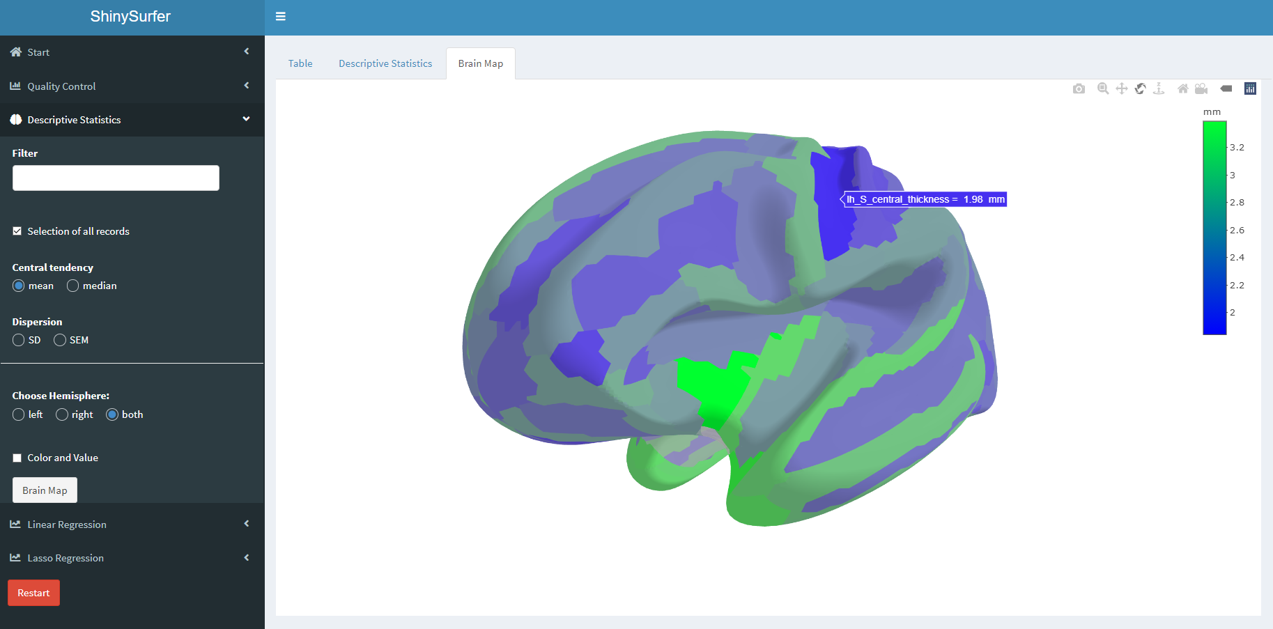
The cyto- and myeloarchitecture of the cerebral cortex differs between cortical areas, reflecting distinct differences in neural function and connectivity (Amunts & Zilles 2015). High-resolution structural MRI allows the non-invasive assessment of the cerebral cortex in the living human brain using surface-based morphometry, implemented in automated software packages such as FreeSurfer (surfer.nmr.mgh.harvard.edu; Fischl 2012). FreeSurfer is able to parcellate the cortical surface of individual brains into neuroanatomically defined areas based on cortical folding patterns using e.g. the Destrieux atlas with 74 cortical areas per hemisphere (Destrieux et al. 2010). After parcellation, FreeSurfer determines mean cortical thickness, cortical area, cortical volume, and curvature for every cortical area. We present ShinySurfer, a novel tool to visualize cortical parcellations and the related cortical parameters, assist in quality control and perform statistical analyses. In this Shiny App, the desterieux atlas is used and divides the brain into 148 regions, each hemispheres has with 74 regions. The Shiny App can be used for personalized custom 3D brain model generation (with the thickness of the cerebral corte), data screening, Raincloud-Plots generation, statistical data output, multi-format image export, lasso regression analysis, simple linear regression analysis.
MRI (Magnetic resonance imaging) plays an important role in the clinical diagnosis of medicine, including Neuroimaging. The human brain is a complex structure and can be divided into two regions (left and right hemispheres), these two hemispheres are connected. "The human cerebral cortex is a highly folded sheet of neurons the thickness of which varies between 1 and 4.5 mm, with an overall average of approximately 2.5 mm. The thickness of the cortex is of great interest in both normal development as well as a wide variety of neurodegenerative and psychiatric disorders. Changes in the gray matter that makes up the cortical sheet are manifested in normal aging, Alzheimer's disease and other dementias, Huntington's disease, corticobasal degeneration, amyotrophic lateral sclerosis, as well as schizophrenia." (Fischl et al. 2000)